Technology

Discover scalable, instrument-free single cell sequencing technology from Parse Bioscience

Technology Overview


Resources

Explore our collection of resources to learn more about technology and its applications from leading researchers

Resources Overview

Company

Providing researchers single cell sequencing with unprecedented scale and ease

About Parse

The Single Cell Solution Immunology Has Been Waiting For

May 16, 2025
|
8 min read
Updated:May 16, 2025


Single cell technologies are revolutionizing immunology.

The immune system is complex and diverse, consisting of various cell types that function in coordination to detect, respond to, and eliminate pathogens. From innate immune cells such as macrophages and neutrophils to adaptive immune cells like B and T lymphocytes, each subset plays a specific role in maintaining immune homeostasis and defending the body against infections. However, this diversity is challenging for researchers aiming to unravel the molecular mechanisms underlying immune responses.

Unlike traditional bulk sequencing approaches that measure average gene expression across entire cell populations, single cell RNA sequencing (scRNA-seq) provides insights into the unique gene expression profiles of individual cells. This granularity is essential for dissecting complex immune responses, identifying rare but functionally critical cell types, and understanding the intricacies of immune cell differentiation and activation, thus facilitating the discovery of new biomarkers, enhancing the understanding of immune-related diseases, and guiding the development of targeted therapies.

Principles of scRNA-seq

Various scRNA-seq methods are currently employed by the scientific community, but they all share a common foundational principle.

First, the tissue of interest—whether solid or blood—is dissociated into a single cell suspension, ensuring the preparation is free of cell aggregates.

The single cells are then partitioned, with the approach varying by technology. Partitioning can be achieved physically, such as encapsulating cells within aqueous droplets, or through individual barcoding to uniquely identify each cell.

Next, RNA from each cell is captured using poly(T) primers or random hexamers to specifically target mRNA. The RNA is then reverse transcribed into cDNA. To ensure accurate cell identification, unique, cell-specific barcodes are incorporated into the cDNA. The cDNA is subsequently amplified to generate sufficient material for sequencing. Finally, the amplified cDNA libraries are sequenced, enabling detailed transcriptomic analysis.

There are two fundamentally different approaches to isolate the cells and prepare the sequencing libraries.

A common approach is droplet-based scRNA-seq and requires microfluidic devices to encapsulate each fresh, live cell into a nanoliter-sized droplet functioning as the reaction vessel. Within these droplets cells are lysed, RNA is captured, and mRNA is reversed transcribed and tagged with cell specific barcoded beads.

An alternative, more direct approach uses the cells themselves as the reaction vessel. The live cells are first fixed, permeabilized, and seeded into a 96-well plate with well-specific barcodes. During an in situ reverse transcription reaction, barcodes are added to identify the sample. The cells are then pooled and split into new multiple wells, each with a unique barcode. Repeated rounds of pooling and splitting ensures that each cell is uniquely labeled and identifiable when sequencing the cDNA libraries (Figure 1).

Figure 1: In the Evercode™ combinatorial barcoding workflow, single cells undergo 4 rounds of splitting and pooling in 96-well plates, without the use of microfluidic devices.

Common Challenges with Traditional Droplet-Based Single Cell Technologies

Despite the advantages of being relatively high-throughput and capable of capturing a wide range of cell types, droplet-based technologies face challenges when applied to immunology research.

Inefficient Capture of Certain Immune Cell Types

Inefficient capture means that the cell’s RNA was not successfully captured and sequenced. There are a few reasons why this may happen.

Cells may be prone to lysis. Granulocytes (neutrophils, eosinophils, basophils) have a fragile nature and high susceptibility to lysis during droplet encapsulation, therefore they may be difficult to capture. Due to the presence of RNAses, these cells have high RNA degradation rates that further contribute to poor transcript recovery.

Some cell subsets are rare and may be masked by more abundant cell populations. For instance, dendritic cells are a low-abundance subset making up for 1-2% of the peripheral blood mononuclear cells. To capture even a small number of these cells using droplet-based methods, researchers must input a very large number of total cells, which increases cost and data volume.

Additionally, they are prone to clumping due to their size, surface adhesion molecules, and tendency to form cell–cell contacts. This leads to inefficient droplet formation or exclusion, or even clogging the chip, causing them to be underrepresented or entirely missed.

Low RNA content cells add another challenge. Immune subsets such as some tissue resident neutrophils with low RNA content can be challenging to detect due to their reduced RNA levels compared to other immune cells. In droplet-based workflows, these low-RNA cells may fail to generate sufficient signal for library preparation, leading to poor-quality barcodes, sparse gene counts, or complete dropout from the dataset. As a result, these biologically important but transcriptionally quiet cells can be underrepresented or misclassified in downstream analyses.

Cell Size and Droplet Compatibility

A droplet has on average 50-60 𝛍M in diameter. Large cells such as activated macrophages or monocytes may exceed this size range, presenting significant challenges.

They may not properly fit within the droplet or the microfluidic channels leading to low capture rate. Additionally, these cells are more prone to clog the microfluidic device leading to disruption of the workflow. The inability to effectively capture these large cells limits the applicability of droplet-based protocols in comprehensive immunological studies that analyze a heterogeneous population including both small and large cells.

Another size-related issue that can clog microfluidic systems is cell clumping. Certain T cell subsets, monocytes, or the DNA released from lysed or dead cells can promote aggregation and clumping. This aggregation can lead to incorrect barcoding, loss of single cell resolution due to higher doublet/multiplet percentage, and, in severe cases, clogging of the microfluidic channels.

Ambient RNA and Red Blood Cells contamination

Extracellular RNA molecules—freely floating RNA not associated with any intact cell—are RNA originating from lysed or dead cells, broken during sample preparation, processing, or handling. They can also be present due to secreted RNA or cell debris. Higher ambient RNA contamination is a common problem in droplet-based technologies.

Blood samples are prone to high red blood cells (RBC) retention. When RBCs lyse they release free-floating hemoglobin’s RNA that can be encapsulated into a droplet and sequenced with the intracellular RNA, leading to cell type misidentification and erroneous gene profiling conclusions. Moreover, as ambient RNA is sequenced as well, a portion of the reads will be wasted on non-useful information the user is still paying for. (Figure 2).

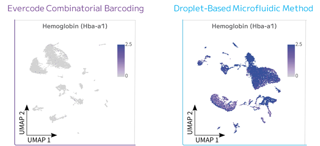
Figure 2: Clustering and Hemoglobin Expression Comparison. Expression of hemoglobin alpha is shown for both Evercode combinatorial barcoding and droplet-based technologies.

Combinatorial Barcoding: An Alternative Approach

Evercode™ combinatorial barcoding has emerged as a robust alternative to droplet-based techniques, especially when applied to immunology research. Unlike droplet-based methods, there is no microfluidic encapsulation, thereby avoiding cell loss associated with droplet formation. The results are a significant increase in sensitivity and differentially expressed genes.

Sensitivity for Fragile Cells

Working without the physical encapsulation of cells into droplets is an advantage when studying with immune cells prone to mechanical stress or lysis. Fragile populations like granulocytes, plasmacytoid dendritic cells, and tissue-resident lymphocytes may rupture during droplet formation due to shear forces. Because combinatorial indexing instead relies on sequential rounds of in situ barcoding, these cells remain intact throughout the workflow, leading to more accurate representation in the final dataset.

High Capture Efficiency for Low-Abundance Cells

Rare immune cell populations, such as antigen-specific CD8 T cells, regulatory T cells, or specific subsets of dendritic cells, are notoriously difficult to capture using droplet-based methods due to the stochastic nature of droplet loading—a large input is required to achieve sufficient sampling. An option around this bias would be to enrich for the population of interest, making this an hypothesis-driven—not a discovery-driven—experiment. Combinatorial barcoding, on the other hand, avoids this problem entirely. This method enables large sample size, up to 5 million single cells uniquely labeled through combinatorial indexing, ensuring that even ultra-rare populations are not lost or underrepresented (Figure 3), thus increasing overall sensitivity. It is this scalability that enables users to detect even the rarest population of cells.

Figure 3: Gene Detection Comparisons. Median genes detected per cell across different sequencing depths for human PBMCs using both Evercode combinatorial barcoding and droplet-based technologies.

Reduced Bias from Cell Size and Aggregation

Immune cells vary significantly in size, morphology, and adhesive properties. Activated macrophages and monocytes, for instance, can be larger and more adherent, leading to clumping or exclusion in droplet-based workflows. Similarly, certain T cell subsets form aggregates or have sticky surfaces that impair droplet formation or barcode delivery. Bypassing the narrow microfluidic channels makes them unaffected by these physical traits. As a result, the use of combinatorial barcoding provides more consistent sampling across diverse immune cell phenotypes.

Conclusion

As immunology research continues to advance, addressing the limitations of current single cell technologies remains crucial. Combinatorial barcoding offers a promising solution for studies requiring high sensitivity and comprehensive analysis of diverse immune cell types. By mitigating the challenges associated with droplet-based methods, this approach holds significant potential for more accurate and insightful immunological discoveries.

About the Author

Laura Tabellini Pierre

Laura Tabellini Pierre, MSc, is a scientific and technical writer at Parse Biosciences with extensive experience in immunology, encompassing both academic and R&D research.
English